38 cell wall diagram with labels
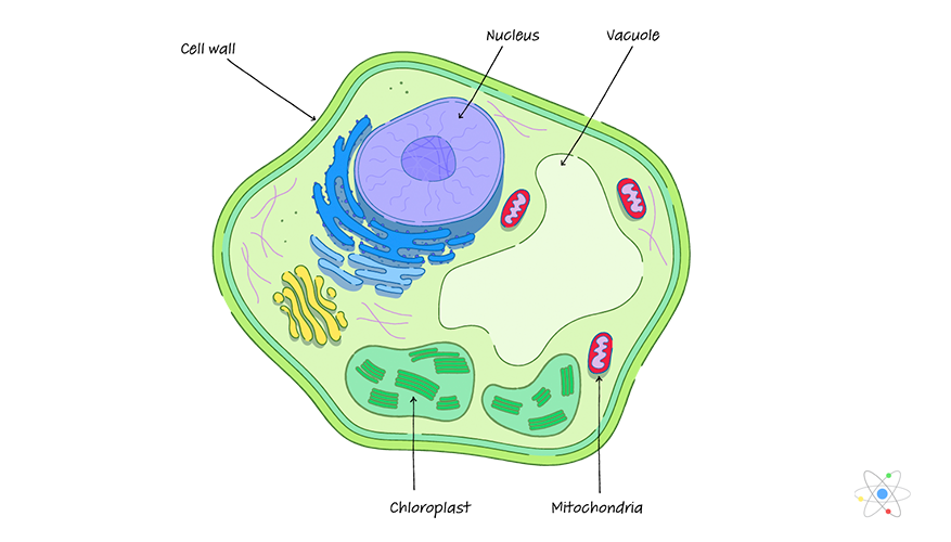
Plant Cell Diagram | Science Trends A plant cell diagram, like the one above, shows each part of the plant cell including the chloroplast, cell wall, plasma membrane, nucleus, mitochondria, ribosomes, etc. A plant cell diagram is a great way to learn the different components of the cell for your upcoming exam. Plants are able to do something animals can't: photosynthesize. Plant Cell - Definition, Structure, Function, Diagram & Types Cell Wall It is a rigid layer which is composed of polysaccharides cellulose, pectin and hemicellulose. It is located outside the cell membrane. It also comprises glycoproteins and polymers such as lignin, cutin, or suberin. The primary function of the cell wall is to protect and provide structural support to the cell.
Gram Positive Bacteria Cell Wall Label Diagram | Quizlet Start studying Gram Positive Bacteria Cell Wall Label. Learn vocabulary, terms, and more with flashcards, games, and other study tools.

Cell wall diagram with labels
Essay Fountain - Custom Essay Writing Service - 24/7 Professional … Professional academic writers. Our global writing staff includes experienced ENL & ESL academic writers in a variety of disciplines. This lets us find the most appropriate writer for … Label Cell Parts | Plant & Animal Cell Activity ... Student Instructions Create a cell diagram with each part of plant and animal cells labeled. Include descriptions of what each organelle does. Click "Start Assignment". Find diagrams of a plant and an animal cell in the Science tab. Using arrows and Textables, label each part of the cell and describe its function. Onion Epidermal Cell Labeled Diagram - schematron.org The epidermal cells of onions provide a protective layer against viruses and fungi that may harm the sensitive tissues. Because of their simple structure and.onion epidermal cell labeled onion cell diagram Wide collections of all kinds of labels pictures online. Make your work easier by using a label. Happy Labeling!
Cell wall diagram with labels. PDF Plant Cell Diagram - Edraw Soft Plant Cell Golgi vesicles Golgi apparatus Ribosome Smooth ER(no ribosomes) Nucleolus Nucleus Rough ER(endoplasmic reticulum) Large central vacuole Amyloplast(star ch grain) Cell wall Cell membrane Chloroplast Vacuole membrane Raphide crystal Mitochondrion Druse crystal Cytoplasm Chapter 25 Flashcards | Quizlet Drag the labels onto the diagram to identify the major renal processes and associated nephron structures. ... What type of receptors embedded in the urinary bladder wall initiate the micturition reflex? decreasing; external ... _____ are hardened cell fragments formed in the distal convoluted tubules and collecting ducts and flushed out of the ... Structure of Cell Wall (With Diagram) - Biology Discussion Cells with secondary wall consist of five layers a three layered secondary wall, the primary wall and the middle lamella. In some cells, such as primary xylem, the secondary thickening materials are laid down in such a way that various patterns are formed on the cell wall, e.g. annular, spiral, reticulate, scalariform and pitted. Prokaryotic cell to label - Labelled diagram - Wordwall Prokaryotic cell to label - Labelled diagram nucleoid region, pili, ribosomes, flagellum, plasmid, cytoplasm, plasma membrane, cell wall, capsule. Prokaryotic cell to label Share by Susanmgillen KS5 Biology Like Edit Content More Leaderboard Log in required Theme Log in required Options Switch template Interactives
Animal Cells: Labelled Diagram, Definitions, and Structure Plant Cells Diagram Plant Cells Organelles and Functions Animals Cells Vs Plant Cells Plant and animal cells have several differences such as plant cells have a cell wall or chloroplasts, but animal cells do not have either. Plant cells are fixed, rectangular in shapes, but animal cells are mostly round and irregular in shape. AS Biology A (Salters-Nuffield) - Edexcel (ii) The student also compared the thickness of the aorta wall of this heart with the thickness of the aorta wall in a giraffe. The thickness of the aorta wall in this heart is 3 mm and in a giraffe it is 15 mm. Give one reason why the aorta wall in a giraffe is much thicker. (1) Animal Cell Labelling Activity | Primary Resources - Twinkl If your students find the Animal Cell Labelling Worksheet useful, this Plant Cell Diagram is a similar labelling activity for plant cells. For something a little more challenging, this Parts of a Cell Cut and Stick Worksheet also challenges students to recall the different components of both animal and plant cells. 03 Label the Cell Diagram | Quizlet Start studying 03 Label the Cell. Learn vocabulary, terms, and more with flashcards, games, and other study tools.
PDF Plant Anatomy: Images and diagrams to explain concepts A diagram of a prokaryotic cell. It lacks organelles and is much smaller and simpler. (LadyofHats Mariana Ruiz. Public Domain). PLANT ANATOMY AND PHYSIOLOGY: IMAGES AND DIAGRAMS TO EXPLAIN 7. 1.2 CELL WALL The cell wall is initially deposited on the surface of the middle lamella. This primary cell wall occurs on the surface of all plant cells ... Bacteria Labeled Stock Illustrations - Dreamstime Salmonella enteri bacteria cell cutaway labeled with flagelli, pili, nucleoid DNA, ribosomes, and cell wall. Science Escherichia coli bacteria E. coli. Medically accurate 3D illustration, labeled. Bacteria shapes, structure and diagram - Jotscroll The bacteria shapes, structure, and labeled diagrams are discussed below. Sizes The sizes of bacteria cells that can infect human beings range from 0.1 to 10 micrometers. Some larger types of bacteria such as the rickettsias, mycoplasmas, and chlamydias have similar sizes as the largest types of viruses, the poxviruses. Cell Wall Structure with Plant Cellular Parts Description ... Royalty-Free Vector Cell wall structure with plant cellular parts description outline diagram. Labeled educational model components description with hemicellulose, pectin and cellulose microfibril vector illustration. cell wall, plant cell wall structure, vector illustration, components description, vector, wall, educational, diagram, cell,
Plant Cell: Diagram, Types and Functions - Embibe Exams Plant Cell Wall It is a rigid layer that is composed of cellulose, glycoproteins, lignin, pectin and hemicellulose. It is located outside the cell membrane and is completely permeable. The primary function of a plant cell wall is to protect the cell against mechanical stress and to provide a definite form and structure to the cell.
Looking at the Structure of Cells in the Microscope A typical animal cell is 10–20 μm in diameter, which is about one-fifth the size of the smallest particle visible to the naked eye. It was not until good light microscopes became available in the early part of the nineteenth century that all plant and animal tissues were discovered to be aggregates of individual cells. This discovery, proposed as the cell doctrine by Schleiden and …
Cell: Structure and Functions (With Diagram) Eukaryotic Cells: 1. Eukaryotes are sophisticated cells with a well defined nucleus and cell organelles. 2. The cells are comparatively larger in size (10-100 μm). 3. Unicellular to multicellular in nature and evolved ~1 billion years ago. 4. The cell membrane is semipermeable and flexible. 5. These cells reproduce both asexually and sexually.
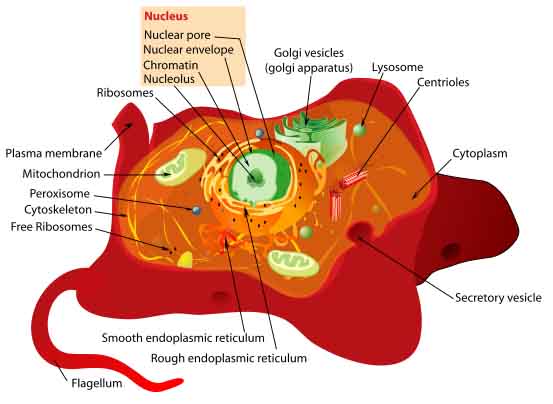
Animal Cell Diagram with Label and Explanation: Cell ... Animal cell is a typical Eukaryotic cell enclosed by a plasma membrane containing nucleus and organelles which lack cell walls, unlike all other Eukaryotic cells. The typical cell ranges in size between 1-100 micrometers. The lack of cell walls enabled the animal cells to develop a greater diversity of cell types.
PDF Human Cell Diagram, Parts, Pictures, Structure and Functions Human Cell Diagram, Parts, Pictures, Structure and Functions The cell is the basic functional in a human meaning that it is a self-contained and fully operational living entity. Humans are multicellular organisms with various different types of cells that work together to sustain life.
Cell Organelles- Definition, Structure, Functions, Diagram In a plant cell, the cell wall is made up of cellulose, hemicellulose, and proteins while in a fungal cell, it is composed of chitin. A cell wall is multilayered with a middle lamina, a primary cell wall, and a secondary cell wall. The middle lamina contains polysaccharides that provide adhesion and allow binding of the cells to one another.
Labeled Plant Cell With Diagrams | Science Trends The parts of a plant cell include the cell wall, the cell membrane, the cytoskeleton or cytoplasm, the nucleus, the Golgi body, the mitochondria, the peroxisome's, the vacuoles, ribosomes, and the endoplasmic reticulum. Parts Of A Plant Cell The Cell Wall Let's start from the outside and work our way inwards.
Definition, Cell Wall Function, Cell Wall Layers - BYJUS The plant cell wall is generally arranged in 3 layers and composed of carbohydrates, like pectin, cellulose, hemicellulose and other smaller amounts of minerals, which form a network along with structural proteins to form the cell wall. The three major layers are: Primary Cell Wall The Middle Lamella The Secondary Cell Wall
Human Cell Diagram, Parts, Pictures, Structure and Functions Human Cell Diagram, Parts, Pictures, Structure and Functions. The cell is the basic functional in a human meaning that it is a self-contained and fully operational living entity. Humans are multicellular organisms with various different types of cells that work together to sustain life. Other non-cellular components in the body include water ...
Animal Cell Model Diagram Project Parts Structure Labeled ... What Is An Animal Cell Biography Source:- Google.com.pk Animal Cells: * have chloroplasts and use photosynthesis to produce food * have cell wall made of cellulose * A plant cell has plasmodesmata - microscopic channels which traverse the cell walls of the cells * one very large vacuole in the center * are rectangular in shape Animal Cells: * don't have chloroplast * no cell wall (only cell ...
Plant and Animal Cell: Labeled Diagram, Structure ... - Embibe Cell Wall: 1. Non-living, rigid, outer boundary. 2. Made up of cellulose, hemicellulose, pectin, lignin, etc. 3. There are many layers, like the middle layer, primary cell wall in a typical plant cell wall. 4. Fungal cell wall is made up of chitin (not cellulose). 5. Protective and provide shape and size. 6. Found only in plant cells. Plasma ...
Label animal cell - Teaching resources - Wordwall 10000+ results for 'label animal cell'. Label Animal Cell Organelles Labelled diagram. by Britter. Label Plant and Animal Cell Labelled diagram. by Catherine34. Label Animal Cell Organelles Labelled diagram. by Mbauer. Animal Cell Label Labelled diagram. by Taraabbott.
Spirogyra Labelled Diagram Draw a neat diagram of Spirogyra and label the following parts: i. Outermost layer of the cell. ii. Organelle that performs the function of. Each cell of Spirogyra filament is cylindrical and consists of 2 parts: cell wall and protoplast. The cell wall surrounds the protoplast, is protective and consists of.
Plant Cells: Labelled Diagram, Definitions, and Structure The cell wall is made of cellulose and lignin, which are strong and tough compounds. Plant Cells Labelled Plastids and Chloroplasts Plants make their own food through photosynthesis. Plant cells have plastids, which animal cells don't. Plastids are organelles used to make and store needed compounds. Chloroplasts are the most important of plastids.
Onion Epidermal Cell Labeled Diagram - schematron.org The epidermal cells of onions provide a protective layer against viruses and fungi that may harm the sensitive tissues. Because of their simple structure and.onion epidermal cell labeled onion cell diagram Wide collections of all kinds of labels pictures online. Make your work easier by using a label. Happy Labeling!
Label Cell Parts | Plant & Animal Cell Activity ... Student Instructions Create a cell diagram with each part of plant and animal cells labeled. Include descriptions of what each organelle does. Click "Start Assignment". Find diagrams of a plant and an animal cell in the Science tab. Using arrows and Textables, label each part of the cell and describe its function.
Essay Fountain - Custom Essay Writing Service - 24/7 Professional … Professional academic writers. Our global writing staff includes experienced ENL & ESL academic writers in a variety of disciplines. This lets us find the most appropriate writer for …


/plasma_membrane-58a617c53df78c345b5efb37.jpg)


Post a Comment for "38 cell wall diagram with labels"